Home
/ Surface Anatomy Of Ribs - Questions on the surface anatomy of the thorax - YouTube : Surface anatomy (also called superficial anatomy and visual anatomy) is the study of the external features of the body of an animal.1 in birds this is termed topography.
Surface Anatomy Of Ribs - Questions on the surface anatomy of the thorax - YouTube : Surface anatomy (also called superficial anatomy and visual anatomy) is the study of the external features of the body of an animal.1 in birds this is termed topography.
Surface Anatomy Of Ribs - Questions on the surface anatomy of the thorax - YouTube : Surface anatomy (also called superficial anatomy and visual anatomy) is the study of the external features of the body of an animal.1 in birds this is termed topography.. Surface anatomy deals with anatomical features that can be studied by sight, without dissection. Costae) are long, flat, curved bones that form the rib cage. Surface markings of the thorax. Rib fractures usually result from blows or from crushing injuries. Illustrations in anterior and posterior view of male torso and back, allowing the lines and regions used in surface anatomy to be displayed (midclavicular line, midline, pectoral thoracic skeleton:
Surface anatomy and surface markings bibliographic record list of illustrations subject index. The ribs form the main structure of the thoracic cage protecting the thoracic organs, however their main function is to aid respiration3. Ascending aorta ends and arch of aorta begins. Colour atlas of human anatomy volume 1, 6th edition, trunk, ribs, pg. The rib 1 head is small, rounded.
Surgical Anatomy of the Chest Wall | SpringerLink from media.springernature.com Surface anatomy (also called superficial anatomy and visual anatomy) is the study of the external features of the body of an animal. In vertebrate anatomy, ribs (latin: But this number may be increased by the. We hope you will use this picture in the study and. Rib anatomy landmarks lungs and ribs anatomy rib anatomy numbers 10th rib anatomy floating ribs anatomy thorax surface anatomy 1st rib anatomy lower rib anatomy human anatomy rib cage muscles rib cage structure typical rib anatomy single rib anatomy anterior. The channel provides a pathway. This is visible and palpable. Anterior chest between ribs i and vi & between the sternum and anterior axillary line.
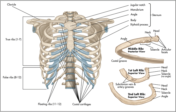
There are twelve pairs of ribs.
Includes images, video, and free quiz. In most tetrapods, ribs surround the chest, enabling the lungs to expand and thus facilitate breathing by expanding the chest cavity. They are twelve in number on either side; Surface anatomy deals with anatomical features that can be studied by sight, without dissection. The rib cage forms the majority of the thoracic skeleton. Lie on top of pectoralis major and tail extends articulation with costal cartilage of rib ii. Learn vocabulary, terms and more with flashcards, games and other study tools. Surface anatomy (also called superficial anatomy and visual anatomy) is the study of the external features of the body of an animal. This muscle assists the internal intercostal muscles. Each rib articulates posteriorly with two thoracic vertebrae by the costovertebral joint. The ribs are elastic arches of bone, which form a large part of the thoracic skeleton. The second rib articulates with the sternum at the sternal angle, making this site an excellent landmark for determining rib number. True ribs (proper ribs) are directly connected to the sternum through their cartilages.
They are twelve in number on either side; We think this is the most useful anatomy picture that you need. Illustrations in anterior and posterior view of male torso and back, allowing the lines and regions used in surface anatomy to be displayed (midclavicular line, midline, pectoral thoracic skeleton: Costae) are the long curved bones which form the rib cage, part of the axial skeleton. Feel for the spinous process of the 7th cervical vertebra (vertebra prominence).
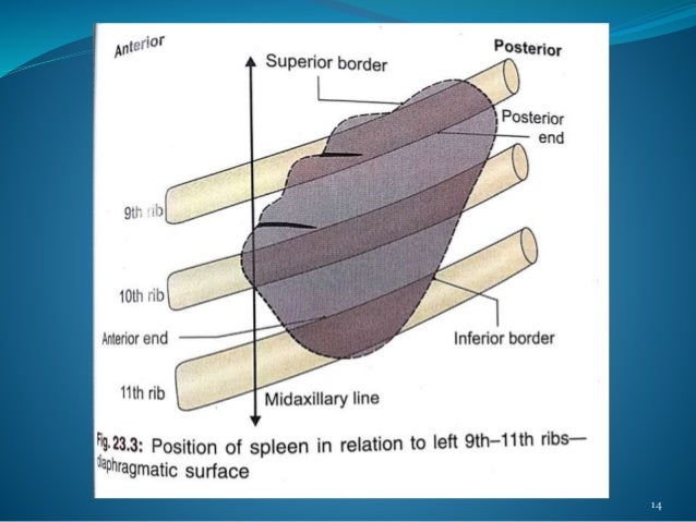
ANATOMY OF SPLEEN AND IT'S APPLIED ASPECT from image.slidesharecdn.com An exception to this rule is those closest to the skin's surface run from the back of the vertebrae to the scapula eg trapezius , rhomboid s, latissimus dorsi , others wrap around the. This image added by anatomy is the amazing science. Ascending aorta ends and arch of aorta begins. Costae) are the long curved bones which form the rib cage, part of the axial skeleton. There are twelve pairs of ribs. But this number may be increased by the. Surface anatomy (also called superficial anatomy and visual anatomy) is the study of the external features of the body of an animal. This is visible and palpable.
The ribs help protect vital organs in the thorax such as the heart.
Anatomy ▶ thorax ▶ bones and cartilages ▶ the ribs. This image added by anatomy is the amazing science. Typical ribs have a normalized general structure, while atypical ribs have slight there are two small grooves in the upper surface of the first rib that house the subclavian vein, nerve, and artery. The ribs form the main structure of the thoracic cage protecting the thoracic organs, however their main function is to aid respiration3. If the rib is set on the incorrect side, then only its anterior end. There are twelve pairs of ribs. We hope you will use this picture in the study and. The loose segment of the wall moves. The exceptions are the 11th and 12th ribs that don't have this surface, which enables them much higher mobility. The rib cage is made up of 12 pairs of ribs, each having a posterolateral bony and an anterior costal cartilaginous component ( fig 4.2 ). Now notice the rib belongs to the side on which it is both ends touch the surface. The ribs/costal cartilages have various attachments to the sternum. Rib anatomy landmarks lungs and ribs anatomy rib anatomy numbers 10th rib anatomy floating ribs anatomy thorax surface anatomy 1st rib anatomy lower rib anatomy human anatomy rib cage muscles rib cage structure typical rib anatomy single rib anatomy anterior.
Colour atlas of human anatomy volume 1, 6th edition, trunk, ribs, pg. In vertebrate anatomy, ribs (latin: Note that the origin of the right oblique fissure is normally at a lower level than. The rib cage is made up of 12 pairs of ribs, each having a posterolateral bony and an anterior costal cartilaginous component ( fig 4.2 ). Counting of ribs in the back:
Locating the Kidneys from i1.wp.com Patterns of bony anatomy of the thoracic cavity and rib cage in anterior and posterior view. In birds this is termed topography. Surface anatomy (also called superficial anatomy and visual anatomy) is the study of the external features of the body of an animal. This image added by anatomy is the amazing science. Anterior chest between ribs i and vi & between the sternum and anterior axillary line. Bony landmarks.—the second costal cartilage corresponding to the sternal angle is so readily found that it is used as a the influence of the obliquity of the ribs on horizontal levels in the thorax is well shown by the following line. (subclavian means below the clavicle. Landmarks of the thoracic wall.
But this number may be increased by the.
Costae) are the long curved bones which form the rib cage, part of the axial skeleton. Learn the true ribs, false ribs, and floating ribs, as well as the difference between in this anatomy lesson, i'm going to cover the rib bones, also called costae in latin. The loose segment of the wall moves. Surface anatomy (also called superficial anatomy and visual anatomy) is the study of the external features of the body of an animal.1 in birds this is termed topography. Patterns of bony anatomy of the thoracic cavity and rib cage in anterior and posterior view. The recipient surface anatomy of a bony defect is typically irregular in its size and shape, which presents the clinician with a challenge as it pertains to grafting. Includes images, video, and free quiz. Typical ribs have a normalized general structure, while atypical ribs have slight there are two small grooves in the upper surface of the first rib that house the subclavian vein, nerve, and artery. We think this is the most useful anatomy picture that you need. The middle ribs are most commonly fractured. There are twelve pairs of ribs. Landmarks of the thoracic wall. In most tetrapods, ribs surround the chest, enabling the lungs to expand and thus facilitate breathing by expanding the chest cavity.
This muscle assists the internal intercostal muscles anatomy of ribs. The loose segment of the wall moves.